
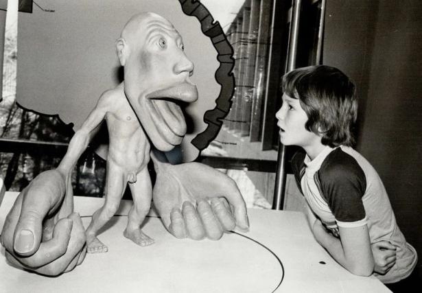
The bizarre-looking ‘homunculus’ is one of neuroscience’s most fundamental diagrams. Found in countless textbooks, it depicts a deformed constellation of body parts mapped onto a narrow strip of the brain, showing the corresponding brain regions that control each part.
But a study published in Nature1 on 19 April reveals that this brain strip, called the primary motor cortex, is much more complex than the famous diagram suggests. It might coordinate complex movements involving multiple muscles through connections to brain regions responsible for critical thinking, maintaining the body’s physiology and planning actions. The new results could help scientists better understand and treat brain injuries.
“This study is very interesting and very important,” says Michael Graziano, a neuroscientist at Princeton University in New Jersey. It’s becoming clear that the primary motor cortex isn’t “just a simple roster of muscles down the brain that control the toes to the tongue”, he says.
Little man in the brain
The idea of the homunculus dates to the late nineteenth century, when researchers noticed that electrically stimulating the primary motor cortex corresponded to specific body parts twitching. Later work found that some body parts, such as the hands, feet and mouth, took up a disproportionate amount of space in the primary motor cortex compared with the rest of the body. In 1937, these findings culminated with the first publication of the motor homunculus, which translates to ‘little man’ in Latin.
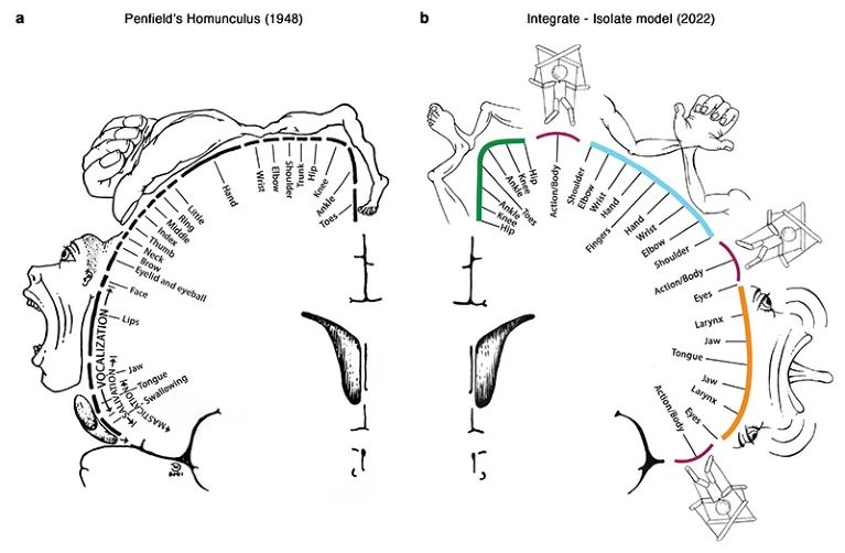
Neurosurgeon Wilder Penfield’s 1948 diagram of the motor homunculus (left) shows the areas of the primary motor cortex that control each body part. A new study redraws the diagram (right), adding regions connected to brain areas responsible for coordinating complex movements.Credit: E. Gordon et al./Nature
As tempting as the idea of a ‘little man inside the brain’ is, Graziano says that more recent evidence suggests the homunculus isn’t completely accurate. For example, a 2002 study2 that he co-authored found that stimulating the primary motor cortex of monkeys caused the animals to carry out coordinated movements rather than simple muscle twitches.
And Angela Sirigu, a neuroscientist at the Institute of Cognitive Science Marc Jeannerod in Lyon, France, and her colleague proposed a new theory in 20083, based on data from people whose arms had been amputated above the elbow. They suggested that there are two systems in the primary motor cortex: one for motor commands and another for muscle synergies that enable coordinated movement.
Quality glimpse
In the latest study, Nico Dosenbach, a neuroscientist at Washington University in St. Louis, Missouri, and his colleagues used functional magnetic resonance imaging (fMRI), which measures the changes in blood flow that occur with brain activity, to scan individuals while they were either resting or moving different parts of their body. The researchers devoted hours of scanning time to each person, which afforded a high-quality glimpse of the primary motor cortex.
Some aspects of the original motor homunculus checked out: there were separate regions that controlled the movements of the foot, hand and face. But the researchers also found three areas interspersed between these regions that were strongly connected with each other and connected to other parts of the brain responsible for goal-driven action planning and other tasks such as regulating blood pressure and pain. These interspersed regions were not specific to any one body part or movement and were activated during action planning.
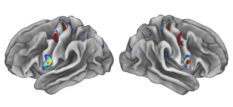
Three coloured spots on each half of the brain show areas that were strongly connected to other regions responsible for goal-driven action planning and tasks such as regulating blood pressure and pain. The hotter the colour, the denser the connections.Credit: Evan Gordon/Washington University
The findings weren’t a fluke: the researchers found the same regions lighting up in large data sets from previously scanned individuals. And when they scanned children, they found that a newborn hadn’t developed this brain network yet, whereas an 11-month-old and a 9-year-old had, supporting the theory that this network coordinates complex action plans, because newborns aren’t yet able to control their movements precisely, Dosenbach says.
The study represents a prime example of the power of fMRI, says Michael Fox, a neurologist at Brigham and Women’s Hospital in Boston, Massachusetts. “We had no idea this secret system of brain regions was there hiding in plain sight until functional brain imaging came along,” he says.
Targets for therapy
The findings could lead to changes in therapy for disorders of the primary motor cortex caused by stroke or injury. Many of the brain areas currently targeted for neurostimulation, a technique used to rehabilitate people with movement disorders, came from trial and error, Fox says. The new homunculus could help explain how and why certain targets work, and identify new or better targets.
Understanding how people recover from damage to the primary motor cortex is really important, says Dylan Edwards, a specialist in neurorehabilitation at the Moss Rehabilitation Research Institute in Philadelphia, Pennsylvania. These findings could help “tailor treatments that might be aligned with specific patterns of deficits”, he says.
Given how long researchers have been probing the motor cortex, “we thought we knew everything about this region”, says Sirigu. “But its organization is much more complex than we have traditionally thought.”
doi: https://doi.org/10.1038/d41586-023-01312-6
References
-
Gordon, E. M. et al. Nature https://doi.org/10.1038/s41586-023-05964-2 (2023).
-
Graziano, M. S. A., Taylor, C. S. R. & Moore, T. Neuron 34, 841–851 (2002).
-
Reilly, K. T. & Sirigu, A. Neuroscientist 14, 195–200 (2008).
Max Kozlov writes for Nature as a life-sciences reporter. His work has also appeared in The Atlantic, Quanta Magazine, The Scientist, The St. Louis Post-Dispatch, Behavioral Scientist, and The Public’s Radio. In his free time, he loves to rock climb, book last-minute travel, and devour just about every journalistic piece he comes across.
Nature is the foremost international weekly scientific journal in the world and is the flagship journal for Nature Portfolio. It publishes the finest peer-reviewed research in all fields of science and technology on the basis of its originality, importance, interdisciplinary interest, timeliness, accessibility, elegance and surprising conclusions. Nature publishes landmark papers, award winning news, leading comment and expert opinion on important, topical scientific news and events that enable readers to share the latest discoveries in science and evolve the discussion amongst the global scientific community.
Spread the word