
The ebb and flow of vehicles along congested highways was what first drew Lisa Manning to her preferred corner of physics, during the summer before she began her graduate studies at the University of California, Santa Barbara. She was mesmerized by the emergent behaviors in the traffic flow — “how you could start out with local rules between cars and get waves of jams through traffic,” she said. But it wasn’t until after she had earned her doctorate in physics in 2008 that Manning started applying that enthusiasm to problems in biology.
During her postdoctoral fellowship at Princeton University, she learned about what’s known as the differential adhesion hypothesis, an idea developed in the 1960s to explain how groups of cells in embryos move and sort themselves out from one another in response to considerations like surface tension. “It was amazing that such a simple physical idea could explain so much biological data, given how complicated biology is,” said Manning, who is now an associate professor of physics at Syracuse University. “That work really convinced me that there could be a place for this kind of thinking in biology.”
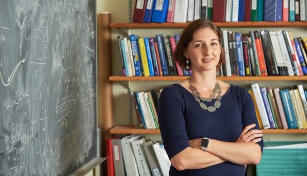
She took inspiration from the dynamics of glasses, those disordered solid materials that resemble fluids in their behavior. Manning found that the tissues in our bodies behave in many of the same ways. As a result, with insights gleaned from the physics of glasses, she has been able to model the mechanics of cellular interactions in tissues, and uncover their relevance to development and disease. Although still at an early point in her career, Manning has won numerous awards for her research, as well as for her teaching and efforts to include and support women in scientific, technical and medical fields. She’s “a rising star,” in the words of one of her colleagues. (Editor’s note: Manning’s work is funded in part by the Simons Foundation, which publishes Quanta.)
Quanta Magazine recently spoke with Manning about how groups of cells move and make transitions between fluid and solid states, how they maintain their boundaries as organs form during embryonic development, and how a breakdown of that process might lead to diseases like cancer. The interview has been condensed and edited for clarity.
Let’s start with the basics. What is the glass problem?
To turn a fluid into a solid, you can just stick a glass of water in the freezer until it turns into ice. It’s a fairly easy process for a physicist to understand: The molecules inside a fluid are all jumbled up, and it’s understood that the system becomes rigid, or solid, because those molecules become ordered.
But glassy materials look microscopically identical in the fluid and solid phases. The big mystery of the glass transition — a mystery we’ve grappled with for over 50 years — is how this is possible. Typically, rigidity is associated with symmetry being broken: The atoms in a fluid look identical everywhere, while in a solid state there are special directions associated with the ordered lattice. In the case of glasses, it’s not clear which symmetry is breaking or how rigidity comes about.
You’ve spoken about how the glass problem has a lot of analogues and applications in fields ranging from artificial intelligence and brain networks to protein folding and morphogenesis. Yet what you just described seems so far removed from something like artificial intelligence. How are those two related?
In glassy systems, we think that many of these interesting properties occur because there’s what’s called a complex potential energy landscape. If you consider the total energy of the entire system as a function of where the atoms are, then in a glass, which is disordered, that landscape is incredibly complex.
It turns out that the neural networks used for deep learning and optimization share a surprisingly large number of properties with glasses. You can think of the nodes of the network as particles, and the connections between them as the bonds between particles. If you do, the neural networks and the glasses have complex potential energy landscapes with nearly identical properties. For example, questions about the energy barriers between states in a neural network are related to questions about how likely it is for a glassy material to flow. So the hope is that understanding some of the properties of glasses can help you understand optimization in these neural networks, too.
Is it typical for materials science to be so relevant to other fields?
Glass physics is peculiar in that it has so many applications. There’s an older connection, going back to the 1980s, between glasses and protein folding. And now it shows up all over the place: in patterns in evolution, in magnets, in the dynamics of social networks. I would say that’s because glasses are a very simple model for a system that is extremely disordered. All of these are networks of nodes, where the connections between nodes are disordered.
So how do you formulate questions about embryogenesis, about organ formation during development, through the lens of this physics problem?
What’s striking is that during development — especially during the early stages, when different layers begin to form in the embryo — cells have to flow over one another for relatively long distances. But then, during later stages of development and as an adult, an animal has to behave more like a solid to support walking and motion.
That means groups of cells have to execute a genetic program fairly regularly to go from being fluidlike, where the cells are all jumbled up and move past one another easily, to a system where they’re locked in place. Meanwhile, you can see the opposite, where fluidization of the tissue might happen, in wound healing, where cells have to move to close an injury, or in cancer, where cells have to move away from a tumor to metastasize. The instructions for all this are in the DNA, at the single-cell level. So what do single cells do to change the global mechanical properties of a whole bunch of cells at the tissue level?

Click here to view video. Lisa Manning, a physicist at Syracuse University, describes how the physics of glassy materials helps to explain how some organs assume their correct shape during embryonic development. Jennifer May for Quanta Magazine
The models for glass transitions have typically been based on molecules or particles, meaning that interactions depend on how far apart, say, one atom is from another. But we’re interested in confluent embryonic tissues, where “confluent” means that there are no gaps or overlaps between the cells. And that means we’re not changing any of the variables we typically associate with a fluid-solid transition, like temperature or how densely the particles are packed. How do you get a fluid-solid transition in a system that doesn’t have any of these properties?
We took an existing model, called a vertex model, which imagines a tight packing of cells in two dimensions as a tiling of polygons, where each vertex moves in response to forces such as surface tension. We used this model to examine properties such as the energy barriers between physical states, or how difficult it was for a cell to move. Those properties in the tissue system showed the hallmarks of what you’d see in a typical glass transition in ordinary materials.
What insights into development have you gained from studying that transition?
We’d like to understand how organs form in development, because if they form improperly, that leads to congenital disease. One hypothesis we have is that some organs actively move through a tissue as they form. In a paper that we just posted on arxiv.org, we discovered that the drag forces — the mechanical fluidlike forces exerted on an organ as it moves — can be sufficient to change the shapes of the cells in ways that help the organ be functional. The fact that the organ is moving through a material that’s either more fluidlike or more solidlike can actually help the organ form properly and do its job. In this paper, we looked at the organ in zebra fish that organizes their left-right asymmetry and helps put, say, their heart on the correct side of the body. We’re really excited about that result, because it suggests that these material properties of the embryo can play a fine-tuned role in helping it develop properly.
So this fluid-solid transition is important in laying out the body asymmetrically?
Even though we all look symmetric on the outside, we’re fairly asymmetric on the inside: The heart is on one side, the liver on the other, and so on. In all vertebrates, that’s driven by a ciliated organ of asymmetry that forms in the early embryo. In this organ, a bunch of cilia stick into a water-filled cavity. The cilia beat in a certain direction and set up a fluid flow inside, and that fluid flow is read out to generate left-right patterning. The direction of flow inside this water-filled organ tells your whole body how to be correctly patterned. That’s why people who have dysfunctional cilia can have inversion symmetry, where different parts of their body end up on the wrong side.
Now, what I’m really interested in is cell shape, because it seems to govern a lot of the physics in these confluent tissues. And it turns out that cell shape plays an important role in this symmetry-breaking organ. In zebra fish embryos, there’s a programmed set of shape changes — possibly governed by fluid-solid transitions — that have to happen to generate this left-right symmetry breaking. For example, the cells at the top of the organ need to get long and skinny, while those at the bottom get short and squat. That way, many more cilia end up on top where they can create a really strong flow.
Beyond asymmetry in development, you’ve also done work on how developing tissues establish and maintain sharp, distinct boundaries. Why is that important?
There are many cases in biology where you want to have a sharp interface, in the sense that the width of the interface is much thinner than a cell diameter. It’s not wiggly. A sharp boundary ensures that you don’t get any mixing between two cell types, which is critical in the segregation of tissues during embryonic development, when cells have to separate and compartmentalize into the gut and the liver and so on.
If you were to look closely at a mixture of two fluids that seem to have a very sharp interface — like a droplet of oil in water — there’s actually a large range over which those molecules mix together, since molecules are so small. But the objects that make up a tissue are cells, and they can be large relative to the scale of an organ. That means these interfaces have to be a lot sharper. If they weren’t so sharp — if they were as mixed as they are at typical interfaces between two fluids — you’d be in deep trouble. And so, if you look at the interfaces that show up in a developing system under a microscope, they’re incredibly sharp.
What makes them so sharp?
Usually, there is a direct relationship between how sharp an interface is and how much surface tension it has. But people have measured surface tension between two cell types — how difficult it is to compress the groups of cells — and those numbers are not that big. The interfaces were orders of magnitude too sharp. Why was there this big difference between the mechanical measurement of surface tension and the sharpness of the interface? We found that it had to do with the cell types being confluent — with the fact that the cells had no spaces between them, and their shape was determined by how adhesive or “sticky” they were.
In such systems, interactions don’t depend on density or how far away your neighbor is, but instead on who your neighbors are and how many neighbors you have. That’s called a topological interaction. Postdoctoral associate Daniel Sussman and I decided to model this system, and together with other faculty at Syracuse we found that these incredibly sharp interfaces were a special feature of systems with topological interactions. That was really exciting and totally unexpected. Moreover, remember that the vertex model we’re using doesn’t only apply to biological cells. So if you wanted a very sharp interface in some kind of artificial structure, such as a foam or a system of self-organizing droplets, you could potentially design materials that have this topological interaction.
If these sharp interfaces are important for maintaining the integrity of tissues, then do we see them break down in diseases?
That’s part of our working hypothesis. We want to know: What holds cancer cells back from metastasizing? The standard take is that they’re held back by a physical barrier called a basement membrane that separates the lining of hollow tissues from the underlying layers. But in many carcinomas (a particular kind of cancer), it’s been seen that cells can break through the basement membrane, and yet they do not move away from the tumor. A few research groups have suggested that the surface tension of a cancer tumor, which is at play in these sharp and soft interfaces, might play an important part in determining whether those cancer cells can escape. How cancer tumors undergo a fluid-solid transition may also play a role in this.
One of our coolest predictions lets us simply look at the shapes of cells to determine whether a tissue behaves like a fluid or a solid, and whether or not those cells can migrate. Our vertex models predicted that in the solid phase, the perimeter of a cell divided by the square root of its area would be exactly 3.81, and that it should rise from 3.81 the more fluid the tissue got. Which is really a crazily strong prediction for biology!
But in 2015, we published a paper with Jeff Fredberg’s group at the Harvard School of Public Health that shows this is exactly right. Since then, my biology colleagues have been a lot more willing to listen to me.
A lot of our work in the past year or so has been geared toward understanding the very deep reason for why that works. It points to an underlying geometric minimal surface problem. Consider the following math problem: You have to tile space with a certain number of tiles, and each tile has to have the same area. What’s the minimal perimeter that all those objects can have? And we have evidence in both two dimensions and three dimensions (because there’s an analogous question in three dimensions) that there’s a minimal surface. In two dimensions, that minimal perimeter-to-area ratio is 3.81. Which means it’s extremely generic: In our model, we may have used a special energy function to understand rigidity, but this implies that any energy function that drives you toward minimizing the perimeter-to-area ratio will give these results. That’s why it’s cool.
On the clinical front, since our research has suggested that cell shapes are really important, we hope to analyze cell shapes from embryos or cancer patients to one day diagnose disease.
We’ve talked a lot about how mechanical forces drive development, structure and disease. What about genetics?
Right. Developmental biologists are also trying to understand how genes and gradients of the signaling molecules called morphogens generate body structure. Obviously, they’re extremely important. Our approach is complementary to that. It’s driving new hypotheses about how cells might control organ formation, by showing that it’s not just biochemical feedback but also mechanical feedback — that the material properties of the tissue itself could be a very robust mechanism for generating patterns in embryos, and that those properties could be targets for therapies.
Have you been working to integrate your mechanical approach with genetic ones?
To be honest, when I started doing this 10 years ago, I thought I could just look up the mechanics of a tissue in some book, and then work on putting biochemical signaling networks on top of it. But I think we’re only just now at a point where we understand the mechanics well enough to couple it with the signaling in models.
That’s why I’m especially excited about new experimental tools called optogenetic tools, which modify the activation of proteins or signaling molecules by flashing light on cells. You can play locally with both the mechanics and the expression of signaling molecules that way, to see how the two interact to generate patterns in developing tissues. There are so many interesting questions to ask.
Jordana Cepelewicz is a staff writer at Quanta Magazine who covers biology. Her writing about mathematics, neuroscience and other subjects has also appeared inNautilus and Scientific American. Before entering the world of science reporting, Jordana did editorial work at Harper’s Magazine, Politicoand Tea Leaf Nation. She graduated from Yale University in 2015 with bachelor’s degrees in mathematics and comparative literature.
Spread the word